NINeTY PERCENT SENSITIVITY.
100 PERCENT COMPASSION.
Women's Imaging
They’re not just images. They’re hope.
Our Women's Imaging department encompasses a fully equipped breast center, pelvic imaging, a fellowship-trained women’s imaging physician and specially trained technologists with over twenty years of experience.
Offering a full range of breast imaging and biopsies at our facilities, allows for continuation of care and more accurate and timely results. We provide screening mammograms, diagnostic mammograms, 3D mammograms, breast ultrasound, breast MRI and ultrasound and MRI guided biopsies.
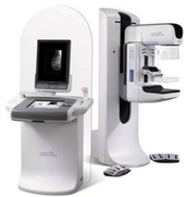
Digital Mammography
Digital Mammography is a regular mammogram where images are acquired using computerized digital technology in place of traditional film.
Digital mammography allows the radiologist to electronically manipulate images where they can be lightened or darkened and examined more closely.
Digital Mammography has been shown to be better at detecting breast cancer in:
- Women who are premenopausal or peri-menopausal
- Women who are under age 50
- Women who have dense breast tissue
Annual Screening Mammography is recommend for all women age 40 and over.
Doctors and scientists agree that early detection is the best defense against breast cancer. If we find cancer in its earliest stage, the chances of surviving it are good. Until now the best way to do that has been with digital mammography.
While digital mammography is still one of the most advanced technologies available, it is only a 2-Dimensional image of the breast.
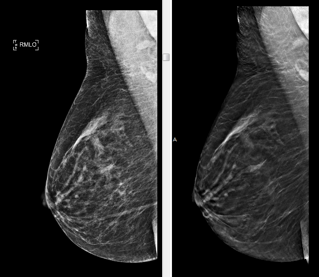
Scientists have developed a new technology called Breast Tomosynthesis, which has been shown in clinical studies to be superior to digital mammography.
Breast tomosynthesis allows doctors to examine breast tissue one layer at a time.
Breast tomosynthesis may be used in conjunction with traditional digital mammography as part of your annual screening mammogram to capture more breast images.
During tomosynthesis images of the breast are taken and then high-powered computing is used to convert digital breast images into a stack of very thin layers or “slices” building a 3-Dimensional mammogram.
Using 3D mammography and digital mammography together for screening has been proven to significantly reduce “call backs” by 20-40%. In addition, 3D mammography finds cancers earlier than 2D mammography alone, with a 27% increase in cancer detection rate and a 40% increase in invasive cancer detection.
What to expect during your exam:
A 3D mammogram is very similar to a traditional mammogram. The technologist will position you, your breast will be placed in compression under a paddle. While in compression the digital mammogram and tomosynthesis images are obtained at the same time. The x-ray arm sweeps in an arch over the breast and multiple images will be taken in seconds. There is no additional compression required with 3D mammography, and it only takes a few seconds longer for each view.
Tomosynthesis / 3D Mammography

Breast Ultrasound
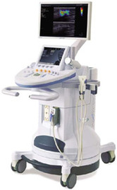
Breast ultrasound is used along with mammography to further evaluate the breast tissue. The ultrasound uses high frequency sound waves to produce images of your breast.
A breast ultrasound will be performed if there is an abnormality on mammography that needs additional evaluation, if there is a mass or lump that the patient feels, or as an additional tool to screen your breast tissue.
Ultrasound Guided Core Biopsies:
This is a minimally invasive procedure that utilizes ultrasound guidance to accurately biopsy a mass or abnormality. A core needle biopsy uses a hollow needle to remove a cylindrical sample of tissue from the breast. The needle is guided to the correct location using ultrasound . A local anesthetic will be used to numb the skin and breast tissue.
Ultrasound Guided Fine Needle Aspiration
A fine needle biopsy uses a thinner needle to remove cells from the area of concern in the breast tissue or lymph nodes. A local anesthetic is used to the numb the area.
Ultrasound Guided Cyst Aspiration
Utilizing ultrasound guidance a needle is place into the breast cyst and fluid is aspirated from the cyst. A local anesthetic is used to the numb the area.
Breast Biopsies